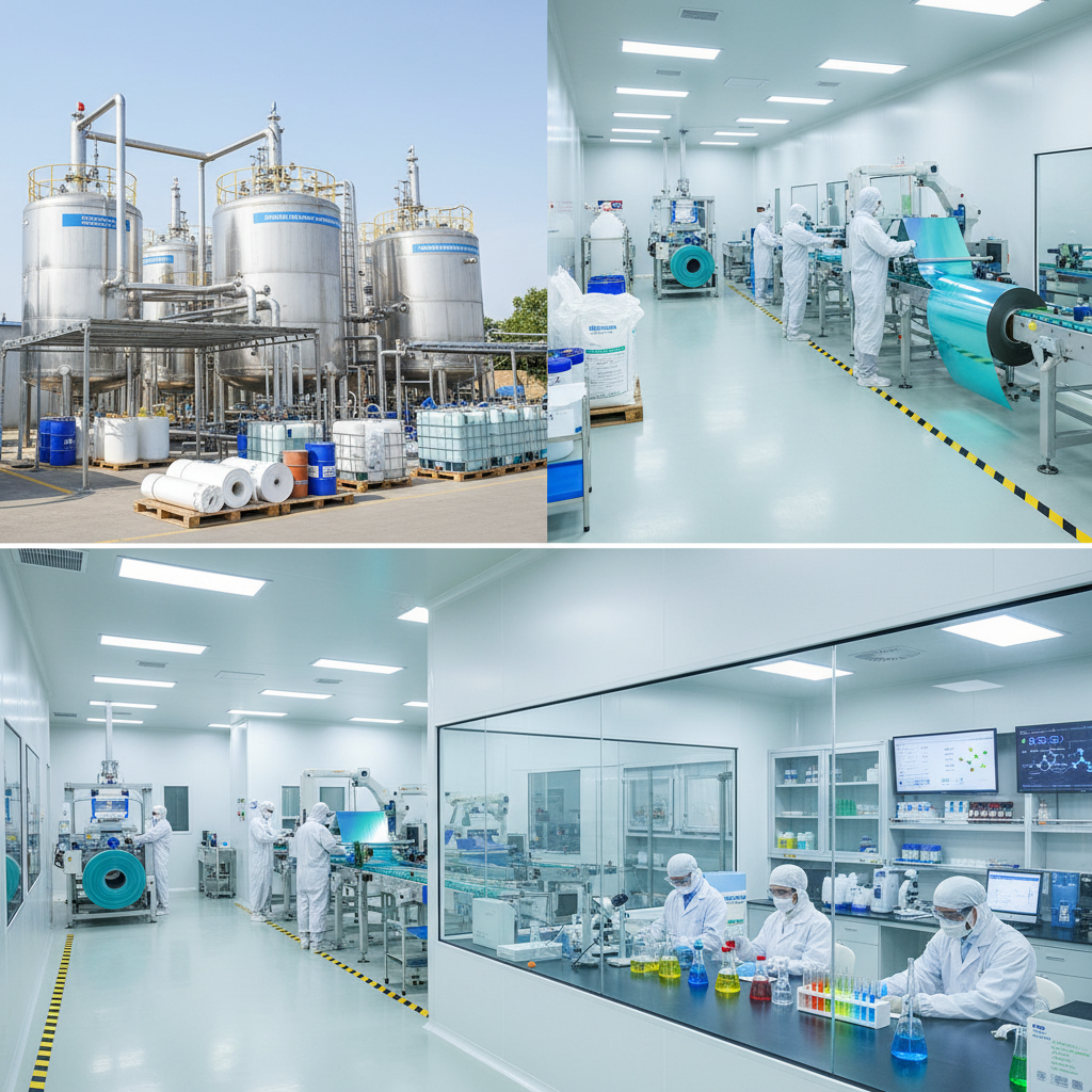
In the precise world of medical diagnostics, radiology films serve as the critical interface between invisible internal anomalies and life-saving medical interventions. Despite the rapid ascent of digital imaging, these specialized materials remain indispensable in various global healthcare settings, providing a permanent, physical record of a patient's physiological state that requires no power source to view.
The global demand for high-quality imaging materials is driven by the need for diagnostic accuracy and archival stability. Whether used in advanced urban hospitals or remote field clinics, the chemical composition and structural integrity of the film determine the clarity of the image, directly impacting the speed and accuracy of a clinician's diagnosis.
Understanding the nuances of radiology films—from their polyester base to the emulsion layers—allows healthcare providers to optimize their imaging workflows. By balancing cost, durability, and image resolution, the industry continues to evolve, ensuring that medical imaging remains accessible and reliable across diverse socioeconomic landscapes.
Global Relevance of Radiology Films
On a global scale, the accessibility of diagnostic imaging is a cornerstone of public health. According to World Health Organization (WHO) guidelines, the ability to perform basic X-ray diagnostics is essential for managing trauma and infectious diseases in low-resource settings. radiology films bridge the gap where digital infrastructure—such as stable electricity and high-cost servers—is unavailable, ensuring that patient care is not dictated by geography.
However, the industry faces the challenge of maintaining strict ISO standards for image quality while reducing the environmental footprint of chemical processing. The shift toward more sustainable, "green" emulsions and recyclable polyester bases is currently a primary focus for manufacturers striving to maintain the efficacy of traditional imaging while adhering to modern environmental regulations.
Defining Modern Radiology Films
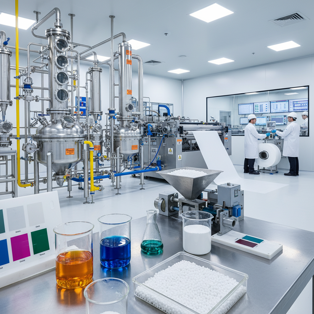
At its most basic level, radiology films are specialized plastic sheets coated with a light-sensitive emulsion, typically composed of silver halide crystals suspended in gelatin. When exposed to X-rays or laser light (in the case of computed radiography), these crystals undergo a chemical change that, after processing, creates a high-contrast image of the internal structures of the human body or industrial components.
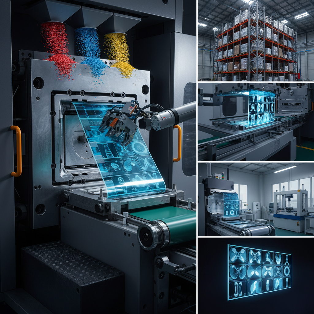
Beyond the chemistry, these films are a product of advanced polymer science. The substrate, usually a high-grade polyethylene terephthalate (PET), must be optically clear, dimensionally stable, and resistant to the heat and chemicals used during the development process. This synergy between chemistry and plastic manufacturing ensures that the final image remains crisp and undistorted over decades of storage.
In the modern industrial context, these materials are not merely "old technology" but are specialized tools for specific humanitarian and professional needs. They provide a tangible, immutable record that is legally recognized in medical malpractice cases and essential for surgical planning in environments where digital screens are impractical.
Core Components of Imaging Materials
The efficacy of radiology films depends heavily on the Optical Density. This refers to the degree of darkness of the film after processing; a high dynamic range in density allows radiologists to distinguish between subtle differences in tissue density, such as detecting a small nodule within a lung field.
Another critical factor is Dimensional Stability. Because these films are often used for measurements (such as measuring the size of a tumor or the alignment of a bone fracture), the plastic base must not shrink or expand. This is achieved through precise thermal treatment during the manufacturing of the plastic substrate to ensure the film maintains its exact scale.
Finally, the Archival Longevity of the material is paramount. To meet international medical standards, radiology films must be resistant to humidity, oxidation, and UV light. This ensures that a patient's medical history can be reviewed ten or twenty years later without the image fading or the film becoming brittle.
Practical Performance Factors
When evaluating the quality of radiology films, professionals look at the balance between sensitivity and contrast. High-sensitivity films require lower radiation doses for the patient, while high-contrast films provide the sharp edges necessary for orthopedic imaging.
The operational cost efficiency is also a major driver. By optimizing the emulsion thickness and improving the efficiency of the developer chemicals, manufacturers can reduce the overall cost per image, making diagnostic services more affordable for clinics in developing regions.
Comparison of Imaging Material Performance Metrics
Global Applications and Use Cases
The application of radiology films extends far beyond the standard hospital X-ray room. In post-disaster relief operations, where power grids are often destroyed, portable X-ray units and physical films allow medical teams to quickly assess fractures and internal injuries in the field, facilitating immediate triage and evacuation.
Furthermore, in the industrial sector, these films are used for Non-Destructive Testing (NDT). Engineers use high-density industrial films to inspect welds in pipelines and aircraft components for microscopic cracks. In these high-stakes environments, the physical film serves as a permanent safety certificate, providing a level of trust and accountability that digital files—which can be edited—sometimes lack.
Long-term Value and Reliability
The tangible benefit of using radiology films lies in their independence. They do not require software updates, proprietary viewers, or cloud access. This "analog reliability" provides a safety net for healthcare systems, ensuring that critical patient data remains accessible even during systemic IT failures or cyber-attacks on hospital databases.
From a psychological perspective, the physical film offers a sense of certainty and dignity in the diagnostic process. For patients in remote areas, holding a physical copy of their scan represents a tangible record of their health journey, which they can take to different specialists without relying on the interoperability of electronic health record (EHR) systems.
Economically, the long-term value is found in the lack of expensive maintenance. While digital systems require constant hardware refreshes and license fees, high-quality archived films, stored in a climate-controlled environment, maintain their diagnostic value for decades with virtually zero ongoing cost.
Future Trends in Imaging Materials
The future of radiology films is being shaped by the push toward sustainability. Researchers are developing biodegradable polymers to replace traditional PET bases, aiming to reduce the plastic waste generated by millions of diagnostic scans globally. This transition toward "Green Radiology" is expected to align the industry with UN Sustainable Development Goals.
Additionally, the integration of hybrid materials is emerging. We are seeing the development of films that can be easily digitized via high-speed scanners while maintaining their physical properties. This ensures a seamless transition between the physical and digital worlds, allowing clinics to enjoy the benefits of both archival stability and digital sharing.
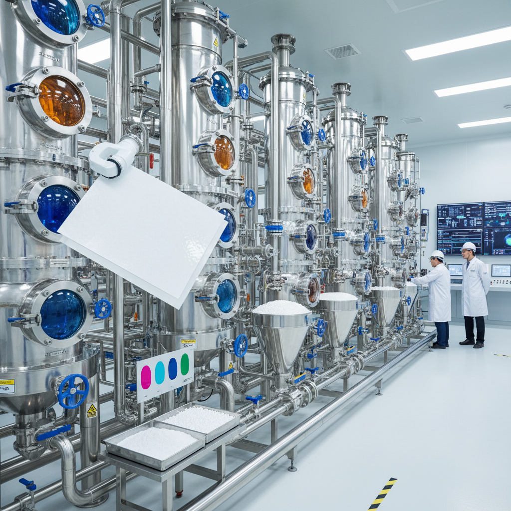
Automation in the production process is also enhancing consistency. AI-driven quality control in the coating process ensures that the emulsion thickness is uniform to the micron level, eliminating "artifacts" in the image and reducing the need for repeat exposures, which ultimately lowers the radiation dose for the patient.
Comparative Analysis of Radiology Film Material Innovations
| Material Type |
Environmental Impact |
Archival Life |
Cost Efficiency |
| Standard PET-Base |
Moderate (Plastic Waste) |
50+ Years |
High |
| Bio-Polymer Base |
Low (Biodegradable) |
20-30 Years |
Medium |
| Silver-Free Emulsion |
Very Low (Non-Toxic) |
30-40 Years |
Medium |
| High-Density Industrial |
Moderate |
100+ Years |
Low |
| Hybrid Digital-Ready |
Moderate |
40+ Years |
High |
| Ultra-Thin Base |
Lower (Less Plastic) |
30+ Years |
Very High |
FAQS
Radiology films are specifically engineered for high X-ray sensitivity and extreme contrast. Unlike photographic film, they use a thicker emulsion layer and a specialized PET base to withstand the chemical processing required for medical-grade clarity and long-term archival stability.
Yes. They are essential for physical archiving, use in low-resource settings without electricity, and for certain industrial NDT applications. They also serve as a legal, immutable backup that cannot be altered by software or deleted by system crashes.
To ensure longevity, films should be stored in a cool, dry environment with low humidity (around 30-50%) and away from direct sunlight. Using acid-free envelopes and avoiding contact with volatile chemicals will prevent the emulsion from degrading.
Artifacts are typically caused by contamination during the processing stage, such as dust on the film, exhausted developer chemicals, or improper rinsing. Using a closed-loop automatic processor significantly reduces these errors compared to manual dipping.
Traditional PET bases are recyclable, but the silver-containing emulsion must be removed first. Many hospitals now use silver-recovery systems to extract precious metals from the waste chemicals, making the process more sustainable.
The choice depends on the body part being imaged. For lungs (low density), a softer contrast film is used. For bones (high density), a high-contrast film is required to see the fine trabecular patterns. Consult the manufacturer's sensitivity chart for specific recommendations.
Conclusion
Throughout this exploration, we have seen that radiology films remain a vital component of global healthcare and industrial safety. By combining precise chemical emulsions with stable polymer substrates, these materials provide a reliable, permanent, and independent method of diagnostic recording that transcends the limitations of digital infrastructure.
Looking forward, the integration of biodegradable materials and AI-optimized manufacturing will ensure that imaging materials continue to evolve without compromising environmental health. For healthcare providers and industrial engineers, investing in high-quality, standardized films is not just a matter of tradition, but a strategic decision to ensure diagnostic accuracy and long-term data security. Visit our website: www.lkintl.com