In the modern landscape of medical diagnostics, the precision of neuroimaging is paramount. The use of high-quality mri brain film serves as a critical bridge between complex digital data and tangible clinical analysis, ensuring that neurologists can examine intricate brain structures with absolute clarity. As healthcare systems shift toward more integrated diagnostic workflows, the demand for stable, high-resolution imaging materials continues to grow globally.
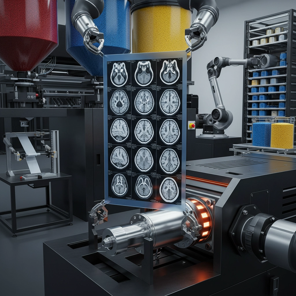
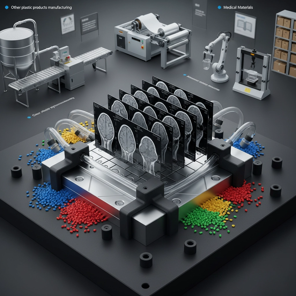
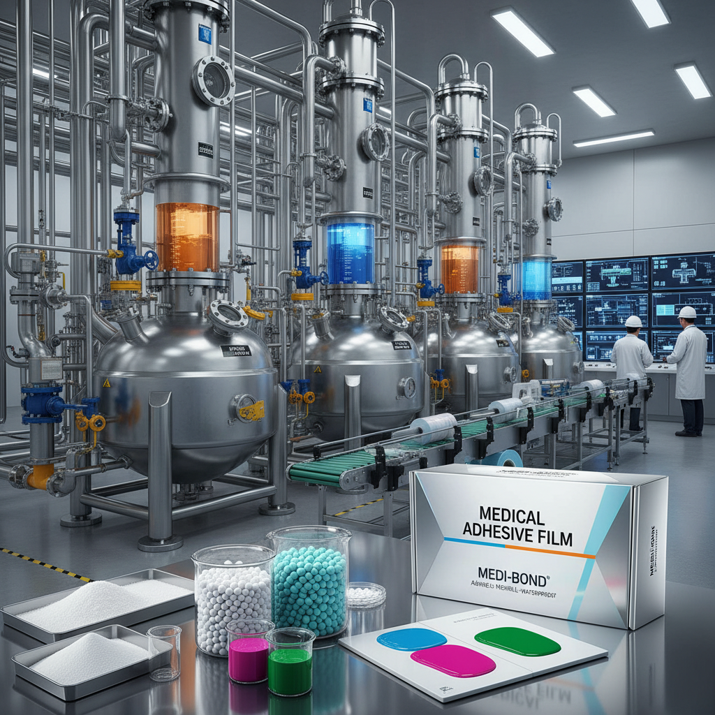
From a manufacturing perspective, the production of these specialized films requires a sophisticated blend of polymer science and optical engineering. The goal is to minimize distortion and maximize contrast, allowing for the early detection of anomalies such as tumors or vascular lesions. By optimizing the chemical composition of the plastic substrates, manufacturers can produce films that are not only durable but also compatible with various imaging modalities.
Understanding the nuances of mri brain film is not merely a technical requirement but a necessity for improving patient outcomes. When the physical medium of the image is flawed, the risk of misdiagnosis increases. Therefore, adopting industry-leading materials ensures that every slice of the brain's architecture is captured and preserved with surgical precision, supporting a higher standard of global medical care.
Global Relevance of MRI Brain Film
The global healthcare burden associated with neurological disorders is rising, with an increasing need for precise diagnostic tools. According to World Health Organization (WHO) trends, the prevalence of neurodegenerative diseases is climbing, making the quality of mri brain film a critical factor in early intervention. In many regions, while digital PACS systems are common, physical films remain indispensable for surgical planning and archival records in remote clinical settings.
The challenge lies in the standardization of these materials. Discrepancies in film density and transparency across different manufacturers can lead to inconsistencies in image interpretation. By adhering to ISO standards for medical imaging materials, the industry aims to eliminate these variables, ensuring that a brain scan produced in one part of the world is interpreted with the same accuracy in another.
Technical Definition and Industrial Meaning
At its core, mri brain film is a specialized medical-grade plastic substrate coated with a photosensitive or thermally reactive layer designed to render Magnetic Resonance Imaging data into a permanent visual format. Unlike standard photographic film, these materials are engineered to handle the specific grayscale gradients required to distinguish between white matter, gray matter, and cerebrospinal fluid in the human brain.
From an industrial standpoint, this product sits at the intersection of the plastics industry and medical technology. The manufacturing process involves high-precision extrusion and coating techniques that prevent "curling" or "yellowing" over time. This stability is vital because medical records must often be stored for decades, necessitating a material that resists oxidative degradation and environmental stress.
The humanitarian significance of this technology cannot be overstated. In developing nations where digital infrastructure may be unstable or unavailable, high-quality physical films allow specialists to transport diagnostic data to remote villages, enabling life-saving surgeries and treatments that would otherwise be impossible without a reliable physical copy of the patient's neuro-anatomy.
Core Components of High-Quality Imaging Film
The durability of mri brain film depends heavily on the choice of the base polymer. Typically, high-grade polyethylene terephthalate (PET) is used to provide the necessary tensile strength and thermal stability, ensuring that the film does not warp when passed through high-temperature printers or scanners.
Optical clarity is the second critical component. For an mri brain film to be effective, the substrate must have a near-zero haze coefficient. This allows the light to pass through without scattering, which ensures that the boundaries of a lesion or the thickness of the cerebral cortex are rendered with sharp, defined edges.
Finally, the chemical interaction between the coating and the substrate ensures long-term adhesion. Advanced primers are applied to prevent the image layer from peeling or flaking, which is essential for films that are frequently handled by multiple medical professionals during a multidisciplinary team (MDT) meeting.
Practical Performance Metrics in Diagnostics
When evaluating the efficacy of imaging materials, clinicians look for specific performance indicators. These include the dynamic range—the ability of the film to capture both the darkest and lightest areas of the brain—and the archival stability, which determines how long the image remains legible without fading.
To provide a clear comparison, we can look at how different formulations of mri brain film perform across various diagnostic requirements. The following data represents a normalized rating of current industry standards.
Comparative Analysis of MRI Brain Film Performance
Global Applications and Use Cases
In high-intensity medical environments, such as trauma centers in North America and Europe, mri brain film is often used for "rapid-review" during emergency neurosurgery. While the primary data is digital, the physical film allows the surgeon to physically map the trajectory of a probe or a shunt against the patient's anatomy in real-time without relying on a screen.
Beyond the operating room, these materials are vital in educational settings. Medical universities worldwide use physical brain films to teach radiology students the art of pattern recognition. By manipulating the film against a light box, students develop a tactile and visual understanding of cranial anatomy that is often lost in purely digital interfaces.
Long-Term Value and Clinical Advantages
The long-term value of investing in premium mri brain film lies in the reduction of diagnostic error. A film with superior contrast ensures that small ischemic strokes or micro-hemorrhages are not overlooked, which can mean the difference between permanent disability and a full recovery for the patient.
Moreover, there is an emotional dimension to this reliability. For patients and their families, the ability to see a physical representation of the brain provides a sense of tangibility and trust. It transforms an abstract digital signal into a concrete piece of evidence, fostering better communication between the physician and the patient.
From a sustainability angle, the industry is moving toward "green" plastic substrates. By utilizing recycled polymers that do not compromise optical purity, manufacturers are reducing the carbon footprint of medical imaging while maintaining the rigid safety and quality standards required for healthcare applications.
Future Trends in Imaging Materials
The future of mri brain film is being shaped by the digital transformation. We are seeing the rise of "hybrid films" that incorporate QR codes or embedded NFC tags. These allow a clinician to scan a physical film and instantly pull up the corresponding 3D volumetric data on a tablet, merging the reliability of plastic with the power of the cloud.
Automation in the coating process is also enhancing consistency. Using AI-driven sensors, manufacturers can now detect microscopic bubbles or thickness variations in real-time, ensuring that every square millimeter of the film provides a uniform response to the imaging process.
As we look toward 2030, the focus will shift toward biodegradable medical plastics. The goal is to create a film that remains stable for the required clinical duration but can be disposed of without contributing to long-term plastic pollution in hospital waste streams.
Comparative Analysis of Future Imaging Material Specifications
| Material Generation |
Optical Transparency |
Environmental Impact |
Clinical Reliability |
| Standard PET Film |
High (85%) |
Moderate |
Stable |
| Low-Haze Polymer |
Very High (95%) |
Moderate |
Excellent |
| Recycled Bio-PET |
High (88%) |
Low |
Stable |
| Hybrid Smart Film |
Very High (94%) |
Moderate |
Superior |
| Degradable Poly-Lactide |
Moderate (80%) |
Very Low |
Short-term |
| Nano-Coated Substrate |
Ultra High (98%) |
Moderate |
Maximum |
FAQS
MRI brain films are specifically designed to render the soft-tissue contrast provided by magnetic resonance imaging, which differs from the high-density contrast needed for X-rays. They prioritize grayscale depth and optical transparency to allow for the visualization of complex brain structures like the hippocampus or basal ganglia, which would be invisible on a standard X-ray film.
Yes, when manufactured using medical-grade PET and high-stability coatings, the film is designed for archival use. To maximize lifespan, they should be stored in a cool, dry environment away from direct sunlight to prevent oxidation and yellowing, ensuring the diagnostic quality remains intact for decades.
Traditionally, these films use petroleum-based plastics. However, the industry is rapidly shifting toward recycled PET and bio-based polymers. Many manufacturers are now producing "green" imaging materials that maintain clinical precision while significantly reducing the environmental footprint of medical waste.
Haze is caused by microscopic imperfections or impurities in the plastic substrate that scatter light. In the context of brain imaging, haze can blur the edges of a tumor or a bleed, potentially leading to a miscalculation of size or location. High-quality films are engineered to be "low-haze" for maximum sharpness.
Most high-grade films are designed for compatibility with standard medical thermal printers. However, it is important to match the film's thermal sensitivity to the printer's heat settings to avoid over-exposure or under-exposure of the image.
Always request the Material Safety Data Sheet (MSDS) and the ISO certification documents from the supplier. Ensure the film meets the specific standards for medical imaging substrates, which guarantee consistent thickness, transparency, and chemical stability.
Conclusion
In summary, the quality of mri brain film is a fundamental pillar of neurological diagnostics. By combining advanced polymer science with a deep understanding of clinical needs, manufacturers can provide a medium that ensures accuracy, durability, and accessibility. From the precision of the base PET layer to the clarity of the final image, every technical detail contributes to the ultimate goal of improving patient care and diagnostic certainty.
Looking forward, the integration of sustainable materials and smart-film technology will continue to evolve, ensuring that imaging remains effective even as the world moves toward a fully digital future. For healthcare providers and procurement specialists, choosing materials that prioritize optical purity and archival stability is an investment in patient safety. Visit our website for more professional solutions: www.lkintl.com